Recent advancements, including detailed imaging and digital resources (like accessible spinal anatomy PDF guides), are revolutionizing our understanding of this complex structure.
Historical Overview of Spinal Study
Early anatomical investigations of the spine date back to ancient Egypt, with the Edwin Smith Papyrus (c. 1600 BC) demonstrating surprisingly accurate observations of spinal injuries. Hippocrates (c. 460-370 BC) also contributed, linking spinal trauma to neurological deficits. However, systematic study truly began with Galen (129-216 AD), whose dissections, though often based on animals, formed the basis of medical knowledge for centuries.
The Renaissance witnessed a surge in anatomical exploration, with Andreas Vesalius (1514-1564) challenging Galen’s inaccuracies through human dissection. Later, figures like Giovanni Battista Morgagni (1682-1771) correlated anatomical findings with clinical symptoms. The 20th and 21st centuries brought revolutionary imaging techniques – X-rays, CT scans, and MRIs – allowing non-invasive visualization of the spine, and the rise of readily available spinal anatomy PDF resources for modern study;
Modern Imaging Techniques in Spinal Anatomy
Revolutionary advancements in imaging have dramatically altered our understanding of spinal anatomy. X-rays, discovered in 1895, provided the first non-invasive view of the vertebral column, though with limited soft tissue detail. Computed Tomography (CT) scans offer cross-sectional images, revealing bony structures with greater clarity. However, Magnetic Resonance Imaging (MRI) stands out, providing exceptional visualization of soft tissues – discs, ligaments, and the spinal cord itself.
Functional MRI (fMRI) assesses spinal cord activity, while myelography, often combined with CT, highlights the spinal cord and nerve roots. These techniques, coupled with 3D reconstruction, enable detailed anatomical study, often accessible through comprehensive spinal anatomy PDF guides, facilitating both clinical diagnosis and surgical planning.
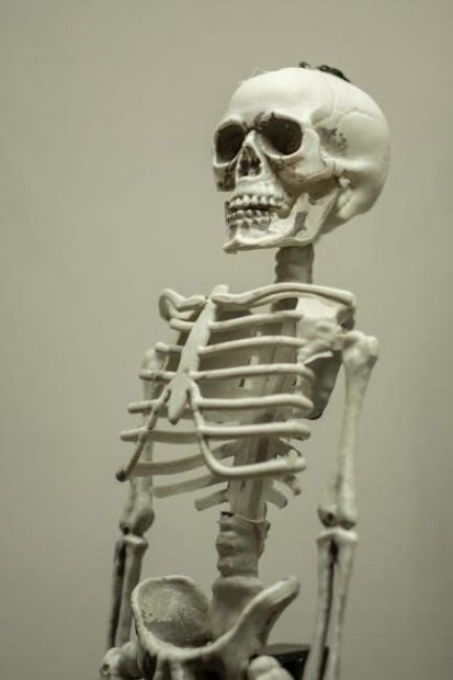
Vertebral Column: Regional Anatomy
The spine is divided into cervical, thoracic, lumbar, sacral, and coccygeal regions, each with unique characteristics detailed in modern spinal anatomy PDF resources.
Cervical Spine – Detailed Structure
The cervical spine, comprising vertebrae C1-C7, exhibits unique features supporting head mobility and protecting the spinal cord. C1 (Atlas) and C2 (Axis) facilitate a wide range of motion. Vertebral bodies are smaller compared to lower regions, reflecting reduced weight-bearing demands. Transverse foramina present in each cervical vertebra allow passage for vertebral arteries, crucial for brain perfusion.
Ligamentous structures, like the anterior and posterior longitudinal ligaments, and the ligamentum flavum, provide stability. Intervertebral discs, composed of annulus fibrosus and nucleus pulposus, act as shock absorbers. Detailed anatomical illustrations, often found in comprehensive spinal anatomy PDF guides, showcase the intricate arrangement of muscles, nerves, and vasculature within this region. Understanding these nuances is vital for clinical assessment and intervention.
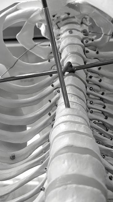
Thoracic Spine – Characteristics and Features
The thoracic spine (T1-T12) is uniquely defined by its articulation with the ribs, providing crucial protection for vital organs. Vertebral bodies progressively increase in size descending down the spine, accommodating greater weight-bearing loads. Costal facets on the vertebral bodies and transverse processes serve as attachment points for the ribs. This region exhibits limited range of motion compared to the cervical or lumbar spine.
Intervertebral discs maintain space and flexibility. Detailed spinal anatomy PDF resources illustrate the complex interplay of muscles, ligaments, and nerves within the thoracic region. Understanding the specific anatomical constraints is essential for diagnosing and managing conditions affecting this area, such as thoracic outlet syndrome or scoliosis.
Lumbar Spine – Key Anatomical Aspects
The lumbar spine (L1-L5) is the lowest portion of the spine, bearing the majority of the body’s weight and enabling significant flexibility. Vertebral bodies are the largest in the spinal column, reflecting this load-bearing responsibility. Facet joints are robust, designed to withstand compressive and rotational forces, but prone to degenerative changes. Intervertebral discs are substantial, providing cushioning and allowing for movement.
Accessing a comprehensive spinal anatomy PDF is crucial for visualizing the intricate arrangement of muscles, ligaments, and nerve roots. Understanding lumbar anatomy is vital for diagnosing conditions like herniated discs, spinal stenosis, and spondylolisthesis, impacting mobility and quality of life.
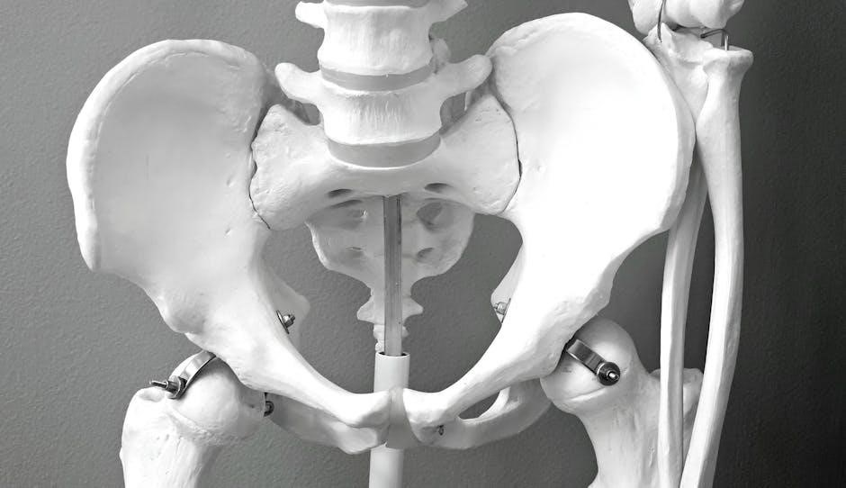
Sacrum and Coccyx – Anatomy and Function
The sacrum, a triangular bone formed by five fused vertebrae, connects the spine to the pelvic girdle, transmitting upper body weight to the lower limbs. Sacroiliac joints, where the sacrum meets the ilium, provide stability but can be a source of pain. The coccyx, or tailbone, consists of typically four fused vertebrae, serving as an attachment point for ligaments and muscles.
Detailed spinal anatomy PDF resources illustrate the complex foramina and canals within the sacrum, crucial for nerve passage. Understanding this region is essential for diagnosing coccydynia and sacral fractures, impacting pelvic stability and neurological function.
Intervertebral Discs and Ligaments
Intervertebral discs provide cushioning, while ligaments stabilize the spine; modern spinal anatomy PDF guides detail their composition and biomechanical roles effectively.
Composition and Function of Intervertebral Discs
Intervertebral discs, crucial components of the spine, are primarily composed of the nucleus pulposus and the annulus fibrosus. The nucleus pulposus, gelatinous in nature, provides hydration and shock absorption, distributing loads effectively. Surrounding it, the annulus fibrosus, a tough, fibrous ring, contains the nucleus and resists tension and torsion.
These discs act as vital spacers, contributing significantly to spinal flexibility and overall height. They enable a range of motion while protecting the vertebrae from impact. Degeneration of these discs, a common age-related process, can lead to pain and neurological symptoms. Detailed spinal anatomy PDF resources illustrate the layered structure and biomechanical function of these essential spinal elements, offering comprehensive insights into their role in maintaining spinal health and stability. Understanding their composition is key to diagnosing and treating related pathologies.
Major Spinal Ligaments – Types and Roles
Spinal ligaments are robust fibrous tissues that provide crucial stability to the vertebral column, limiting excessive motion and preventing injury. Key ligaments include the anterior longitudinal ligament (ALL), preventing spinal flexion, and the posterior longitudinal ligament (PLL), resisting hyperextension. The ligamentum flavum connects adjacent laminae, assisting in maintaining upright posture.
Interspinous and supraspinous ligaments further stabilize the spine, particularly during movement. These ligaments work synergistically to control spinal motion and protect the spinal cord. Comprehensive spinal anatomy PDF guides detail the precise attachments and functional roles of each ligament. Understanding ligamentous anatomy is vital for diagnosing sprains, strains, and other spinal injuries, and for planning effective treatment strategies to restore spinal integrity.

Spinal Cord and Nerve Roots
Detailed PDFs showcase the spinal cord’s intricate organization, transmitting signals via nerve roots, impacting motor and sensory functions throughout the body.
Spinal Cord – Internal Structure and Organization
The spinal cord, a vital component of the central nervous system, exhibits a remarkably organized internal structure; Modern anatomical resources, often available as comprehensive spinal anatomy PDF guides, detail its gray and white matter arrangement. The central ‘H’-shaped gray matter contains neuronal cell bodies, dendrites, and synapses, crucial for processing reflexes and initial sensory input.
Surrounding this is the white matter, composed of myelinated axons forming ascending (sensory) and descending (motor) tracts. These tracts facilitate rapid communication between the brain and peripheral nervous system. Detailed diagrams within these PDFs illustrate the specific organization of these tracts – dorsal columns, lateral corticospinal, and spinothalamic, for example – and their respective functions. Understanding this precise organization is fundamental to diagnosing and treating spinal cord injuries and neurological disorders. The cord is also segmented, with each segment giving rise to paired spinal nerves.
Dermatomes and Myotomes – Clinical Relevance
Dermatomes, areas of skin innervated by specific spinal nerve roots, and myotomes, muscle groups controlled by individual nerve roots, are critical for neurological assessment. Spinal anatomy PDF resources frequently include detailed dermatomal and myotomal maps. Clinically, these maps are invaluable in pinpointing the level of spinal cord injury or nerve root compression.
Weakness or sensory loss in a specific dermatomal or myotomal distribution suggests a lesion at that corresponding spinal level. For instance, weakness in wrist extension (C6 myotome) or numbness in the thumb (C6 dermatome) can indicate a C6 nerve root issue. These concepts are thoroughly explained and visually represented in modern anatomical guides, aiding in accurate diagnosis and targeted treatment planning. Understanding these patterns is essential for effective neurological examination.

Spinal Musculature and Fascia
Detailed spinal anatomy PDF guides showcase the intricate layers of muscles and fascia, vital for stability, movement, and protecting the delicate spinal cord.
Deep Back Muscles – Anatomy and Function
The deep back muscles, often overlooked, play a crucial role in spinal stability and controlled movement. These muscles, including the transversospinales (multifidus, rotatores, and interspinales) and the erector spinae group (iliocostalis, longissimus, and spinalis), are positioned close to the vertebral column.
Modern spinal anatomy PDF resources emphasize their segmental nature, meaning they primarily act on individual vertebrae. The multifidus, for example, provides crucial stabilization, particularly in the lumbar region, preventing excessive motion and injury. The erector spinae, while extending the spine, also contributes to lateral flexion and rotation.
Understanding their attachments, fiber orientation, and synergistic actions is paramount for clinicians. These muscles work continuously to maintain posture and protect the spinal cord, making them essential for functional movement and overall spinal health. Detailed anatomical illustrations within these PDFs aid in visualizing these complex relationships.
Superficial Back Muscles – Anatomy and Function
Superficial back muscles, including the trapezius, latissimus dorsi, and levator scapulae, are larger and more prominent than their deep counterparts. While not directly acting on the vertebral column in the same way, they significantly influence spinal posture and movement through their attachments to the scapula and upper limbs.
Modern spinal anatomy PDF guides highlight their role in gross movements like shoulder elevation (trapezius), arm adduction and extension (latissimus dorsi), and scapular control (levator scapulae). These muscles contribute to spinal stability by controlling scapular position, which impacts the force vectors acting on the spine.
Clinical understanding of these muscles is vital, as dysfunction can lead to postural imbalances and pain. Detailed anatomical charts within these resources demonstrate their origins, insertions, and actions, aiding in accurate diagnosis and targeted rehabilitation strategies.

Vascular Supply of the Spine
Detailed spinal anatomy PDF resources emphasize the vertebral arteries and venous plexus, crucial for spinal cord perfusion and drainage, impacting neurological function.
Arterial Supply to the Vertebral Column and Spinal Cord
The arterial supply to the spine and spinal cord is a complex network vital for maintaining neurological function. Vertebral arteries, originating from the subclavian arteries, ascend through the transverse foramina of the cervical vertebrae, ultimately forming the basilar artery. This basilar artery supplies the posterior circulation of the brain.
Spinal arteries, branching from the vertebral arteries, provide direct blood flow to the spinal cord. These include the anterior and posterior spinal arteries, running along the length of the cord. Radicular arteries, arising from intercostal, subcostal, and lumbar arteries, contribute to the collateral circulation.
Modern spinal anatomy PDF resources highlight the importance of understanding these vascular pathways for surgical planning and diagnosing vascular compromise. Variations in arterial supply are common, and detailed anatomical knowledge is crucial for avoiding iatrogenic injury during spinal procedures.
Venous Drainage of the Spine
Venous drainage from the spine occurs through an intricate network of veins. Vertebral veins accompany the vertebral arteries, ultimately draining into the brachiocephalic veins. Intervertebral veins connect the anterior and posterior venous plexuses, forming a longitudinal pathway. The posterior venous plexus is particularly prominent and receives contributions from the spinal nerve roots.
Anterior venous plexus drains the anterior spinal cord and vertebral bodies. These plexuses communicate with the azygos and hemiazygos systems in the thorax. Understanding venous anatomy is crucial, as epidural venous engorgement can impact spinal cord perfusion.
Contemporary spinal anatomy PDF guides emphasize the clinical relevance of venous drainage patterns, particularly in cases of spinal trauma or tumor compression. Detailed illustrations within these resources aid in visualizing this complex network.

Clinical Correlations & Resources (PDF Focus)
Spinal pathologies, like herniations, are best understood with strong anatomical knowledge; readily available spinal anatomy PDF resources offer detailed visual guidance for clinicians.

Common Spinal Pathologies – Anatomical Basis
Understanding common spinal pathologies necessitates a firm grasp of vertebral anatomy, disc composition, and ligamentous support. Herniated discs, for instance, directly relate to intervertebral disc degeneration and weakening of the annulus fibrosus, impacting nerve root compression. Spinal stenosis, a narrowing of the spinal canal, often stems from age-related changes like facet joint hypertrophy and ligamentum flavum thickening.
Spondylolisthesis, vertebral slippage, is linked to defects in the pars interarticularis. Scoliosis, abnormal spinal curvature, can arise from congenital vertebral malformations or neuromuscular imbalances. Accurate diagnosis and treatment planning rely heavily on detailed anatomical knowledge, often supplemented by resources like comprehensive spinal anatomy PDF guides. These resources visually demonstrate the anatomical basis of each pathology, aiding in precise clinical assessment and surgical intervention.
Where to Find Reliable ‘Spinal Anatomy PDF’ Resources
Locating trustworthy spinal anatomy PDF resources requires careful consideration. University medical libraries often provide access to detailed anatomical atlases and textbooks in digital format. PubMed Central and Google Scholar can yield open-access articles with anatomical illustrations. Reputable publishers like Elsevier and Springer offer premium anatomical resources, sometimes with trial access.
Caution is advised when downloading from unfamiliar websites; prioritize sources with clear authorship and academic affiliations. Many institutions offer online learning platforms with integrated anatomical models and downloadable guides. Always verify the information’s accuracy and currency, as anatomical understanding evolves. Seeking recommendations from medical professionals can also direct you to reliable spinal anatomy PDF materials.